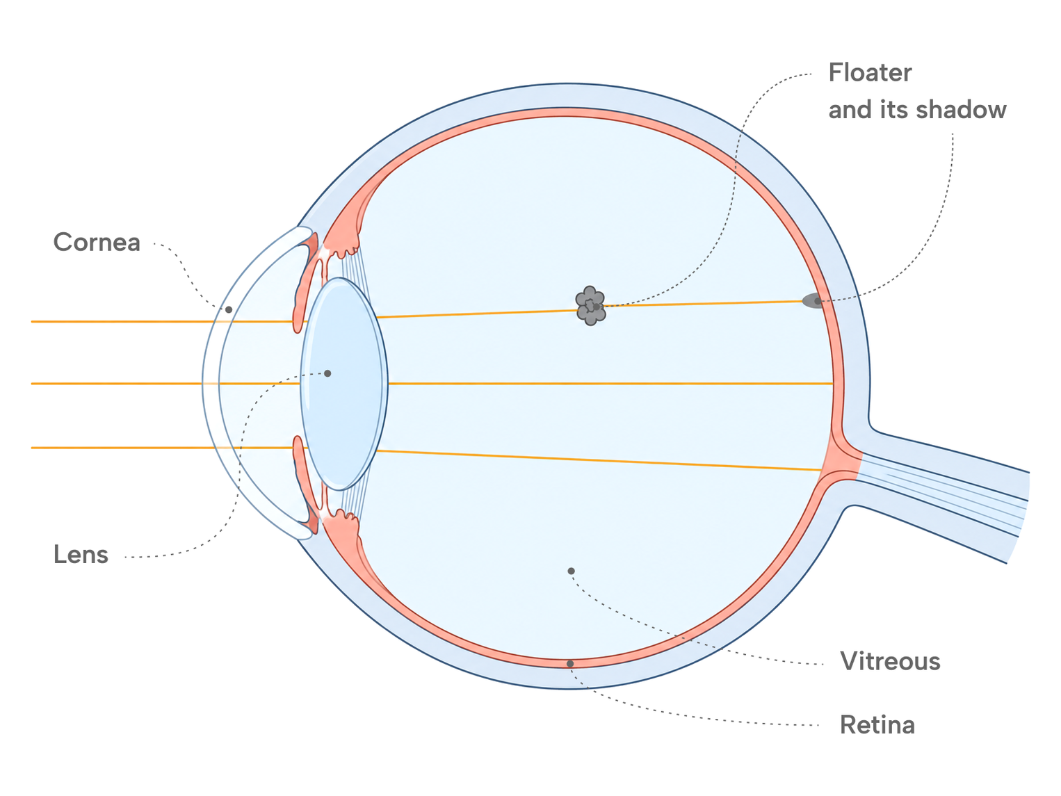
Eye floaters are small shapes that drift across your vision — specks, threads, rings or cobweb-like strands — and move when the eye moves. They are shadows cast on the retina by clumps or strands inside the vitreous, the clear gel that fills the back of the eye. Most floaters come from age-related change in that gel. A small number signal a problem at the retina, and the features that separate the two are specific enough to describe.
Key takeaways
- Floaters are shadows cast on the retina by clumps or strands in the vitreous gel. They move with the eye and are easiest to see against a bright, plain background.
- The most common cause is age-related change in the vitreous, including posterior vitreous detachment (PVD), in which the gel separates from the retina.
- A sudden shower of new floaters, flashes of light, or a shadow spreading across part of the vision needs an eye examination promptly, because these can be signs of a retinal tear or retinal detachment.
- In a large cohort of people examined for new PVD symptoms, 5.4% had a retinal tear and 4.0% a retinal detachment at the first visit. Most had neither.
What are eye floaters?
The back two-thirds of the eye is filled with the vitreous, a clear gel made mostly of water held in a fine scaffold of collagen fibres. Light passes through it on the way to the retina. When fibres in the gel clump together, or when the gel's structure changes, the clumps are no longer fully transparent and cast a faint shadow on the retina. That shadow is the floater.
Floaters appear to drift because the vitreous is not fixed in place. When the eye moves, the gel and the clumps inside it lag slightly behind and then settle, so the shadow glides across the field of view and is hard to look at directly. Floaters are most visible against a uniform bright background — a clear sky, a white wall or a blank screen — where nothing else competes with the shadow.
The medical term for floaters is myodesopsia. Because the shadow is produced inside the eye, floaters are not removed by blinking, rubbing the eye or using eye drops.
What causes eye floaters?
Age-related change in the vitreous
With age the vitreous gradually becomes more liquid and its collagen fibres clump into strands. At some point the gel shrinks enough to separate from the back of the eye. That separation is called posterior vitreous detachment (PVD). Floaters occur more often with PVD, but they also occur without one, and a PVD can be present without the person noticing new floaters. PVD is a common age-related event rather than, by itself, a disease, and it happens in each eye at its own time.
When PVD produces symptoms, floaters are the main one. In a cohort of 8,305 people examined for a new symptomatic PVD, 94.6% reported floaters and just under half also reported flashes of light. Flashes happen when the separating gel tugs on the retina, and they are covered in more detail below because they change what an examination is looking for.
Short-sightedness (myopia)
Vitreous changes tend to happen earlier in myopic eyes. In the same cohort, PVD occurred on average about a year earlier for each dioptre of myopia. Myopia does not change what floaters look like, but it does mean they can appear at a younger age.
Blood or inflammatory cells in the vitreous
Anything that puts cells into the vitreous can produce floaters. Bleeding into the gel — for example from a retinal tear, from abnormal blood vessels in diabetic retinopathy, or after an injury — is seen as a shower of small dots or a haze. Inflammation inside the eye (uveitis) releases inflammatory cells that are also seen as floaters. In these situations the floaters are a symptom of the underlying condition, and the examination is aimed at finding that condition rather than at the floaters themselves.
After eye surgery
PVD can occur earlier in eyes that have had cataract surgery, so new floaters after surgery may reflect vitreous change arriving sooner. They are still assessed in the same way as new floaters in an unoperated eye, because a retinal tear or detachment has to be ruled out.
What is in the vitreous | How floaters tend to appear | |
|---|---|---|
| Age-related vitreous change, including PVD | Clumped collagen strands | Gradually, or as a new floater or group of floaters over days |
| Retinal tear | Blood or pigment cells released when the retina tears | Suddenly, often as many small dots, with or without flashes |
| Bleeding from retinal blood vessels | Blood | Suddenly, as dots or a haze that can affect vision |
| Inflammation inside the eye (uveitis) | Inflammatory cells | Over days, often with other symptoms such as light sensitivity or redness |
Important
Floaters from a retinal tear and floaters from ordinary vitreous change can look identical to the person seeing them. What separates them is the pattern of onset and what an examination of the retina finds, not the appearance of the floater itself.
When do floaters need an eye examination?
A floater that has been present for months and has not changed is not, on its own, a reason for an urgent visit. What matters is change. Three patterns are associated with a retinal tear, which is an opening in the retina created when the separating vitreous pulls hard enough on a spot where it is firmly attached.
A sudden increase in floaters. Many new floaters appearing over hours or a day — often described as a shower, a swarm or a cloud of dots — can be blood or pigment cells released from a tear. This differs from a single new strand that appears and then stays much the same.
Flashes of light. Brief flashes, arcs or streaks, usually at the edge of the vision and more noticeable in the dark, come from the vitreous tugging on the retina. Flashes can occur during a PVD when the vitreous tugs on the retina. New flashes are still a reason for an examination, and the concern is higher when they appear together with a sudden increase in floaters or a shadow in the vision.
A shadow or curtain. A dark area at the edge of the vision that spreads over hours or days, or a sensation that a curtain has come down over part of the view, can mean that the retina has begun to detach. This is the sign that carries the greatest urgency.
These signs are usually in one eye, because PVD and retinal tears happen in one eye at a time. Floaters in one eye are therefore not unusual, and the side is less important than whether the pattern above is present.
The reason these signs are treated with urgency is the numbers behind them. In the 2018 cohort of 8,305 people examined for a new symptomatic PVD, 5.4% had a retinal tear and 4.0% a retinal detachment at the first visit. Among the 7,522 whose first examination found neither, 1.8% went on to develop a tear or detachment within a year; the median interval was 22 days, and 70% of these later events occurred within six weeks. The probability was much higher in eyes with bleeding into the vitreous at the first visit, with an area of thin retina called lattice degeneration, or with a history of a tear or detachment in the other eye. A retinal tear found early is treated with laser or freezing treatment to seal it, which is why the examination is timed to the onset of symptoms rather than left until the floaters become bothersome.
When to see a doctor
Contact an ophthalmologist the same day if you notice a sudden shower of many new floaters, new flashes of light, or a dark shadow or curtain spreading across part of your vision — especially if these appear together or in one eye. These can be signs of a retinal tear or detachment, and the examination needs to happen before a tear has a chance to progress.
What does an eye examination for floaters involve?
The examination looks at the retina rather than at the floaters. Drops are used to widen the pupil (a dilated examination), and the ophthalmologist views the retina through a lens under bright light, paying particular attention to the far periphery where tears from a PVD usually occur. The retina may be examined with gentle pressure on the eyelid to bring the very edge into view. Dilation blurs near vision and increases light sensitivity for several hours, which is worth planning for if you are driving or working on a screen afterwards.
If the retina is intact, the floaters are documented and the visit ends with advice about the changes that would need a return visit. Because some tears appear in the weeks after the first examination, a follow-up examination is often arranged within that period; in the cohort above, 45% of the later tears and detachments were found at a scheduled follow-up rather than because of new symptoms. If a tear is found, it is usually treated at the same visit or soon after.
Do eye floaters go away?
Floaters from vitreous change rarely disappear physically, but they usually become far less noticeable. Two things contribute. The clump can settle lower in the gel, out of the line of sight, and the brain gradually stops registering a shadow that does not change. Many people who were very aware of a new floater in the first weeks find that months later they see it mainly when they look for it against a bright background.
For the small number of people whose floaters remain large, dense and persistently in the line of sight, two procedures exist. Vitrectomy removes the vitreous gel, and the floaters with it, through small incisions; it is a surgical procedure with its own risks, and it is uncommon for floaters alone. Laser vitreolysis uses a laser to break up a floater into smaller fragments without entering the eye. Whether either is considered depends on how much the floater is affecting daily life and on what the examination shows, and that discussion belongs with the ophthalmologist who has examined the eye. Neither procedure is the first step for sudden new floaters, flashes or a curtain-like shadow; those symptoms are examined first to rule out a retinal tear or detachment.
There is no eye drop, supplement or exercise that has been shown to remove floaters, because the clump is inside the gel where none of these reach.
Medical information
This article is general information about eye floaters and is not medical advice. The cause of floaters cannot be determined from their appearance alone, and the signs described here are reasons for an examination rather than a diagnosis. Decisions about examination, follow-up and treatment are made with an ophthalmologist who has examined your eyes.