Bloodshot eyes are common, and most episodes settle without any lasting effect. The white of the eye turns red for several different reasons, and those reasons range from a short night's sleep to a condition that needs to be seen the same day. What separates them is usually not how red the eye looks. It is where the redness sits, and what else is happening alongside it.
Key takeaways
- Redness in the white of the eye comes either from conjunctival blood vessels widening or from blood leaking beneath the conjunctiva, and the two look different from each other.
- A sharply outlined bright red patch with no pain, no discharge and no change in vision is usually a subconjunctival haemorrhage, which usually clears within one to three weeks.
- Allergic conjunctivitis usually affects both eyes at the same time, while epidemic keratoconjunctivitis often begins in one eye and reaches the other several days later.
- Redness forming a ring around the cornea, or redness with eye pain, light sensitivity or changed vision, is assessed by an eye doctor the same day.
What makes eyes look bloodshot
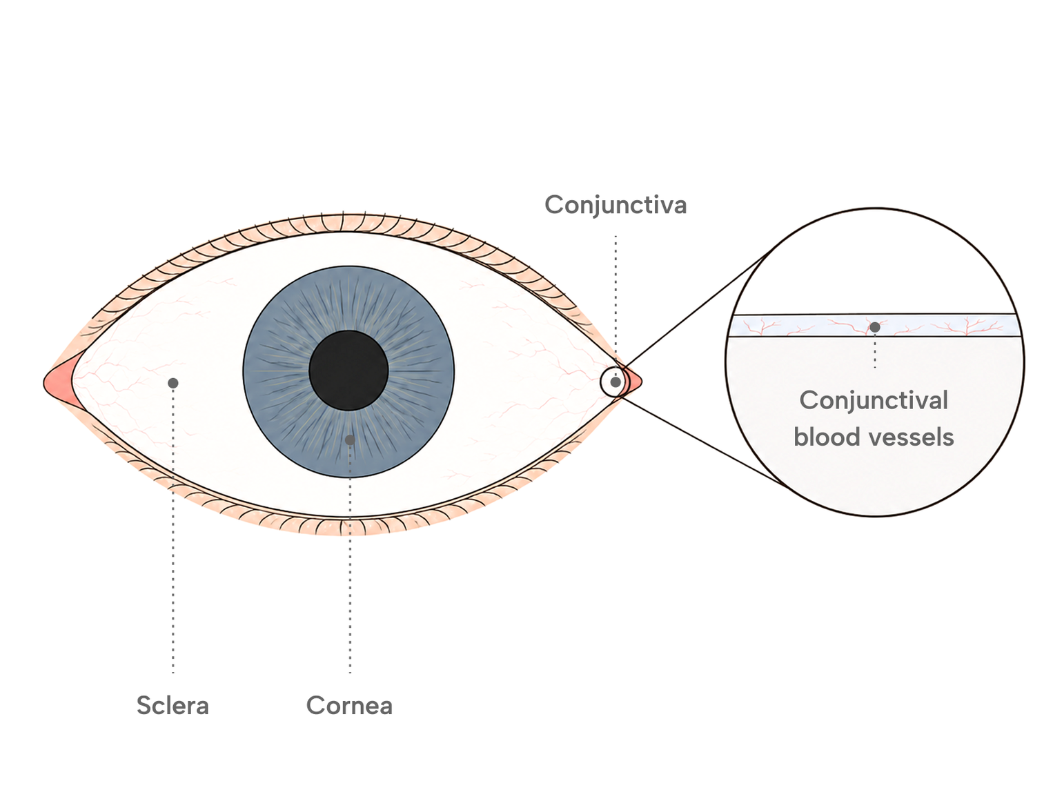
The white of the eye looks bloodshot when blood vessels in the conjunctiva become visible. The conjunctiva is a thin, clear membrane that covers the white of the eye and lines the inside of the eyelids, and it carries a dense network of small vessels. Those vessels can widen, or they can break.
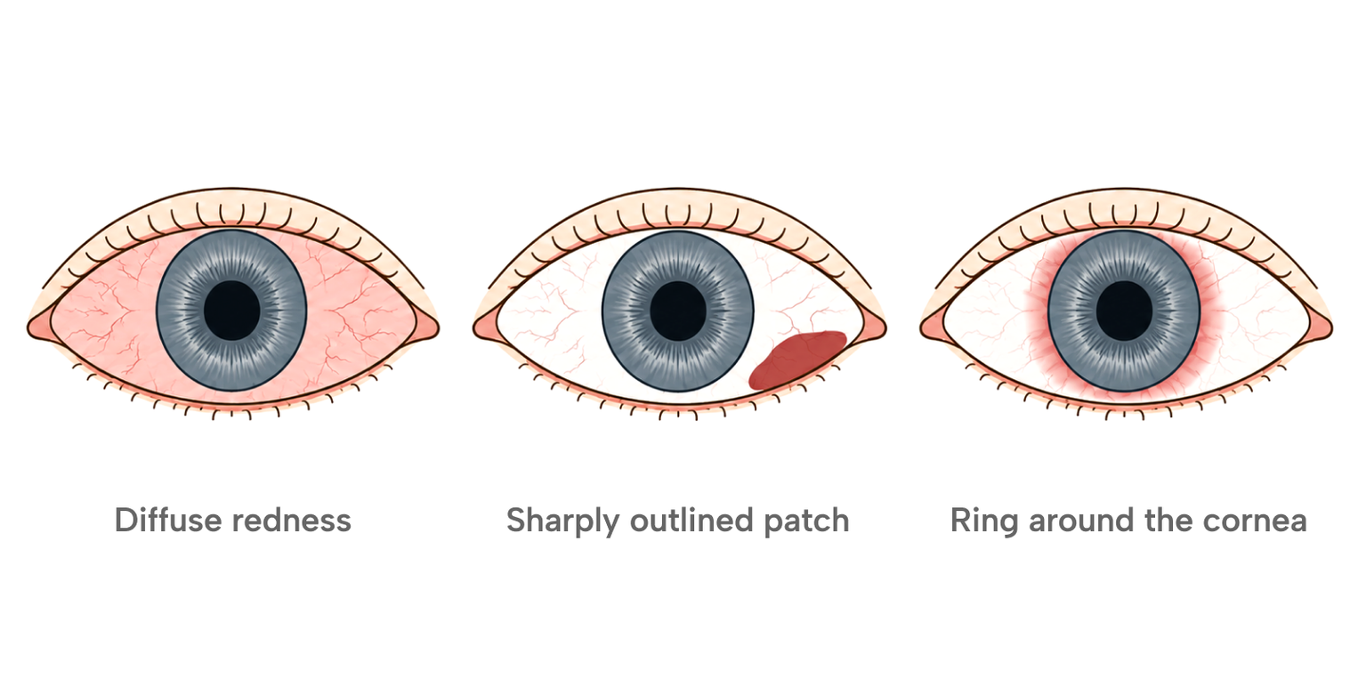
Widening produces a diffuse redness in which individual vessels can still be traced across the surface. A broken vessel produces something different. Blood collects between the conjunctiva and the tissue beneath it, and that area becomes a solid block of red with no separate vessels visible inside it.
Where the redness sits carries information of its own. Redness spread across the surface of the eye is described as conjunctival injection. Redness concentrated in a ring immediately around the cornea is described as ciliary, or circumcorneal, injection. That distribution can point to a problem deeper than surface irritation, particularly when it comes with pain, light sensitivity or reduced vision.
Common causes that are not linked to eye disease
Many episodes of bloodshot eyes come from everyday irritation rather than from a serious eye condition. Dryness, short sleep, long stretches of screen work, airborne allergens, contact lens wear, smoke, chlorinated water and wind all widen conjunctival vessels. Redness from these causes is usually diffuse and arrives without pain or any change in vision.
When the trigger is environmental, such as smoke, wind, dry air or airborne allergens, the redness often reaches both eyes. Contact lens irritation or another local trigger can affect one eye alone, so pain, light sensitivity and blurred vision matter more than whether the redness is one-sided.
Redness of this kind generally settles once the trigger is no longer present. What it does not do is come with eye pain, marked light sensitivity or blurred vision. Those belong to a different group of causes, set out further down.
Contact lens wear belongs on this list with a qualification. A lens sits directly on the ocular surface, and friction, deposits on the lens surface and reduced tear film stability all irritate the conjunctiva. Improper contact lens use is also recognised as a cause of bleeding beneath the conjunctiva, not only of surface irritation. Long-term contact lens wear can cause its own set of problems.
Important
If an eye becomes red while you are wearing contact lenses, take the lenses out and leave them out until the eye has been examined. Redness in a lens wearer that comes with pain, light sensitivity or blurred vision is treated as urgent.
What a bright red patch on the white of the eye means
A bright red patch with a sharp outline, no pain and no change in vision is usually a subconjunctival haemorrhage. Blood from a small conjunctival vessel collects beneath the membrane and cannot spread past it, so it forms a well-defined block of colour against the white of the eye.
A subconjunctival haemorrhage is generally confined to one eye, and it appears most often on the outer lower part of the white. The patch does not fade under pressure and is not accompanied by discharge. Many people only find it when they look in a mirror.
The colour changes over the following days. The patch starts bright red, deepens, and then turns yellow-green as the trapped haemoglobin is broken down, following the same sequence a bruise follows on the skin. Many cases clear in about two weeks, and a range of one to three weeks is commonly described. Larger bleeds, and bleeds in people taking anticoagulant or antiplatelet medication, can take longer.
Why a conjunctival vessel breaks, and what is checked when these bleeds keep returning, is set out separately.
Why only one eye may be red
Whether one eye or both eyes are red narrows the range of likely causes, though that pattern does not identify a cause on its own. Triggers that arrive through the environment, such as allergens, smoke and dry air, usually reach both eyes at once. Causes that begin locally, such as a broken vessel or an injury, usually affect one.
Allergic conjunctivitis is described as affecting both eyes at the same time, because the trigger is environmental. Epidemic keratoconjunctivitis follows a different pattern. It often begins in one eye and spreads to the other several days later, and when both eyes are involved the first one is usually the more severely affected. A subconjunctival haemorrhage is generally one-sided.
One red eye that also feels painful, is sensitive to light, or sees less clearly than the other belongs with the causes set out further down. Blurred vision confined to one eye has its own separate set of causes.
Redness concentrated on the eyelid rather than on the white of the eye is a separate matter. A lump in the lid margin follows a different pattern from redness of the ocular surface.
Redness with discharge, itching, or crusting
Conjunctivitis is inflammation of the conjunctiva, and the type of discharge is one of the more useful ways to tell its forms apart. Viral conjunctivitis typically produces a watery discharge. Bacterial conjunctivitis typically produces a thick, pus-like discharge. Allergic conjunctivitis has itching as its most prominent feature.
Epidemic keratoconjunctivitis is a highly contagious adenoviral infection involving both the conjunctiva and the cornea. Symptoms appear after an incubation period of four to ten days, seven on average. The cornea is typically involved about four days after the conjunctivitis begins, and corneal involvement is one of the features that separates this infection from conjunctivitis that stays on the conjunctival surface. When the cornea is involved, light sensitivity and blurred vision become more important than redness alone.
The infection stays strongly contagious for about two weeks after symptoms begin, and it spreads through eye secretions, hands, towels and shared washing items. Most cases improve within two to three weeks. Severe cases can persist for three to four weeks or longer, and faint corneal opacities may remain afterwards, though these usually clear on their own.
The table below sets out how these causes usually present. It describes patterns rather than a way to diagnose your own eye.
Appearance | What usually comes with it | One eye or both | |
|---|---|---|---|
| Surface irritation | Diffuse redness, individual vessels still traceable | Grittiness; no discharge | Often both; one eye possible with a local trigger |
| Subconjunctival haemorrhage | Sharply outlined solid red patch | No pain, no discharge, vision unchanged | Usually one |
| Viral conjunctivitis | Diffuse redness with swelling | Watery discharge; a tender node in front of the ear | Often one first, then both |
| Bacterial conjunctivitis | Diffuse redness | Thick, pus-like discharge | One or both |
| Allergic conjunctivitis | Diffuse redness with swelling | Itching; watery discharge | Usually both at once |
Redness that needs same-day attention
Some patterns of redness are assessed the same day. A subconjunctival haemorrhage does not cause pain, light sensitivity, reduced visual acuity or an abnormal pupil. When any of those appear alongside a red eye, the cause is something other than a simple surface bleed.
Redness forming a ring around the cornea, rather than spreading across the surface, is the pattern most worth recognising. That distribution is reported alongside conditions that need prompt examination, including inflammation involving the iris or the cornea, acute glaucoma and scleritis.
Redness that follows an injury is also treated differently from redness that appears on its own, because a bleed can accompany damage to deeper structures.
Chemical exposure follows its own timeline. Symptoms can look mild at first and then worsen over the following hours, so the initial level of pain is not a reliable guide to how much damage has been done.
When to see a doctor
See an eye doctor the same day if a red eye comes with eye pain rather than mild irritation, with reduced or blurred vision, with marked sensitivity to light, with a ring of redness around the cornea, or with nausea and haloes around lights. The same applies after an injury, and to any red eye that is painful while contact lenses are being worn. After a chemical splash, rinsing the eye immediately with saline, or with clean running water if saline is not at hand, comes before anything else, and the eye then needs emergency care. Rinse first even if that delays leaving for the hospital, and have the eye examined afterwards even if the pain has eased.
Redness after laser vision correction
Redness in the days after laser vision correction is a recognised finding rather than an unexpected one. Eye surgery appears among the causes of subconjunctival haemorrhage, and a red patch on the white of the eye can follow a procedure. A subconjunctival haemorrhage does not itself affect vision, and it clears along the same course as a bleed from any other cause.
This does not replace the warning signs above. After eye surgery, redness that comes with worsening pain, reduced vision, marked light sensitivity, increasing discharge, or symptoms that are getting worse rather than settling should be assessed promptly rather than treated as part of expected recovery.
What to expect during the first days and weeks is set out separately, as is the question of which symptoms continue beyond the early period.
Medical information
This article is general information about eye redness and is not medical advice. Individual results differ. Decisions about whether a symptom needs examination, and about any treatment, should be made with a qualified ophthalmologist after examination.