ICL corrects short-sightedness by adding a lens inside the eye rather than reshaping the cornea. The lens sits behind the iris and in front of the eye's own lens, where it is not normally felt or visible from outside. Because the cornea is not reshaped, suitability and follow-up centre on measurements inside the front of the eye, as well as on the small incision used to insert the lens.
Key takeaways
- ICL stands for implantable collamer lens. It is placed in the posterior chamber of the eye, behind the iris, and the natural lens stays in place.
- Corneal tissue is not removed, so suitability depends more on measurements inside the front of the eye — including anterior chamber depth, angle anatomy and endothelial cell density — than on corneal thickness alone.
- The U.S. approval covers adults aged 21 to 60 within defined ranges of myopia, together with an anterior chamber depth of 3.00 mm or more and a prescription stable within 0.5 dioptres for a year.
- The lens can be removed or exchanged, and follow-up commonly includes checks of eye pressure, the lens position and the corneal endothelium.
What is ICL surgery?
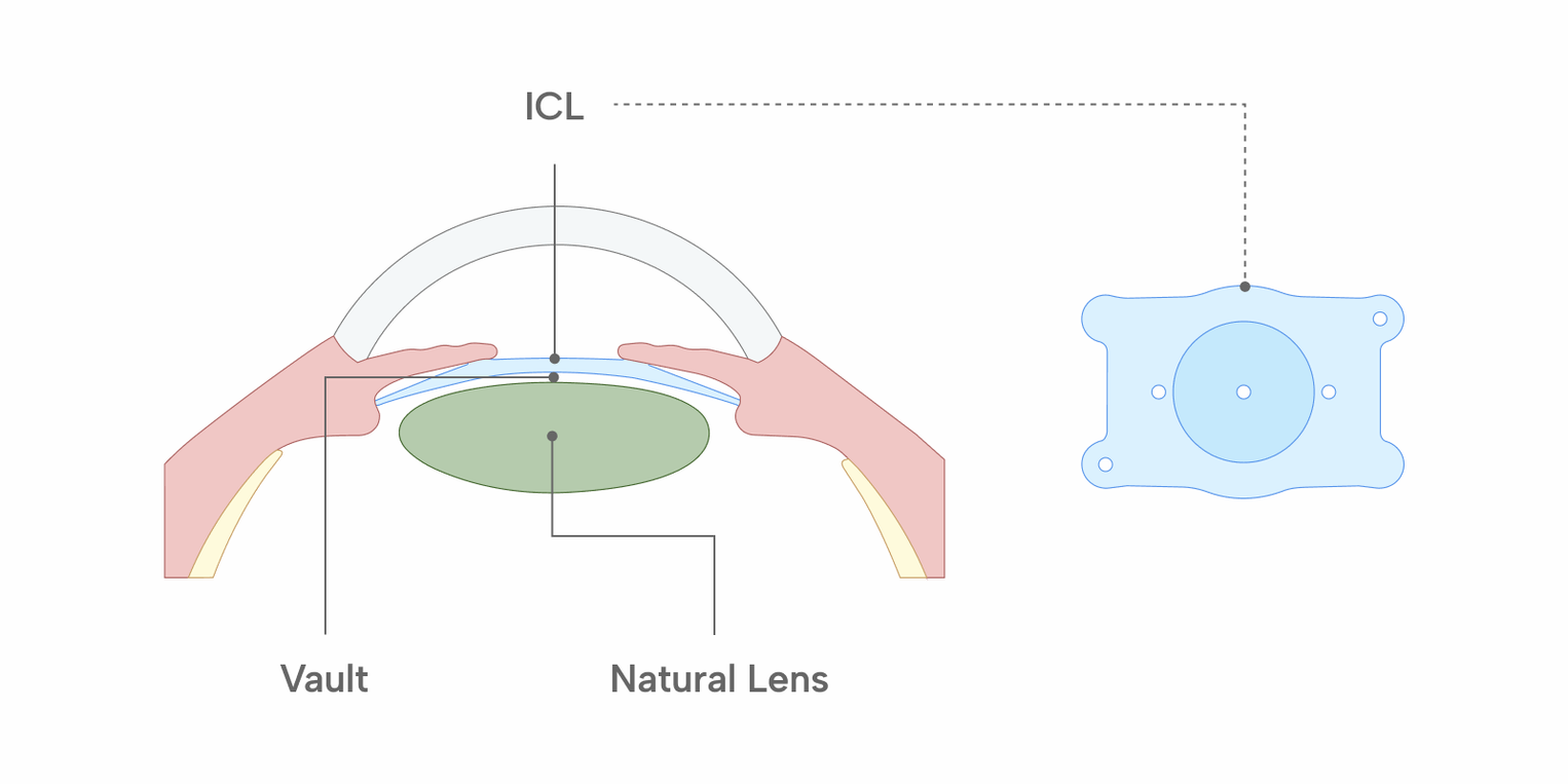
ICL stands for implantable collamer lens. A thin, flexible lens is folded, inserted through a small incision at the edge of the cornea, and positioned in the posterior chamber — behind the iris and in front of the eye's own crystalline lens. The natural lens is not removed. The incision is small, and whether it requires a suture is decided during surgery.
Collamer is the material the lens is made from, a blend of collagen and a polymer. It transmits oxygen and nutrients, and the lens sits directly in front of the eye's own lens, where fluid exchange between the two continues.
The lens is intended to remain in the eye long term rather than to be exchanged at set intervals. It can be removed or exchanged if that becomes necessary, because the eye's own tissue has not been altered to accommodate it.
What is the difference between anterior chamber and posterior chamber lenses?
ICL belongs to a wider group of lenses that are placed in an eye which keeps its own natural lens, known as phakic intraocular lenses. These lenses are classified by where they sit and by what holds them in place. A lens placed in front of the iris, in the space between the cornea and the iris, is an anterior chamber lens. A lens placed behind the iris and in front of the natural lens is a posterior chamber lens. ICL is a posterior chamber lens.
Anterior chamber lenses are grouped by what holds them in position: designs fixated to the iris, and designs supported in the angle where the iris meets the cornea. The corneal endothelium — the cells lining the back of the cornea — is monitored after any phakic lens is implanted, and how close a lens sits to those cells depends on where it is placed.
A posterior chamber lens is not fixated to the iris. Its supports rest in the groove behind the iris, known as the ciliary sulcus, and the lens is made in a range of overall diameters so that one can be chosen to fit the space measured before surgery. The gap it leaves in front of the natural lens is the measurement followed afterwards, because too little gap and too much gap are associated with different problems.
The table below sets out where each type of lens sits, what holds it, and what is measured afterwards. It does not compare outcomes.
Anterior chamber lens | Posterior chamber lens (ICL) | |
|---|---|---|
| Where it sits | In front of the iris, between the cornea and the iris | Behind the iris, in front of the natural lens |
| What holds it in place | Fixated to the iris, or supported in the drainage angle | Supported in the groove behind the iris; not fixated to the iris |
| How the lens is sized | Sizing method varies by design | Chosen from the available overall diameters to fit the space behind the iris |
| What is measured afterwards | Endothelial cells; eye pressure | Vault (gap to the natural lens); eye pressure; endothelial cells |
How is ICL different from laser procedures?
Laser procedures change the shape of the cornea. ICL does not remove corneal tissue and instead adds an optical element inside the eye, reaching it through a small incision at the corneal edge. Both change how light is focused; they do it at different points in the eye's optical path.
Where the correction is placed determines what the pre-operative examination looks for. For a laser procedure the central question is how much corneal tissue there is to work with. For ICL it is how much room there is inside the front of the eye, and whether the cells lining the back of the cornea are healthy enough.
PRK and LASEK, which treat the corneal surface after the epithelium is removed, are covered separately in our guide to PRK and surface ablation.
The table below compares the two approaches on what is changed and what is measured, rather than on outcomes.
Laser procedures | ICL | |
|---|---|---|
| What is changed | The shape of the cornea | A lens is added inside the eye |
| Corneal tissue | Some is removed | Not removed; a small incision is made for insertion |
| Where it acts | At the corneal surface or within it | Behind the iris, in front of the natural lens |
| If adjustment is needed later | Removed tissue does not return | The lens may be removed or exchanged in selected situations |
| Key measurement beforehand | Corneal thickness and shape | Anterior chamber depth and endothelial cell density |
| What is monitored afterwards | Refraction and corneal healing | Eye pressure, lens position and endothelial cells |
What does recovery involve?
Many people notice improved vision within a day or two, and eye drops are prescribed for a period after surgery. The incision is small, so the early restrictions centre on avoiding pressure on the eye and keeping water out of it rather than on protecting a healing corneal surface.
Follow-up in the first days checks eye pressure and the position of the lens. Pressure is monitored in the early period because the lens sits in the path along which fluid produced inside the eye travels toward its drainage channels. If that flow is obstructed, pressure can build behind the iris and push it forward, narrowing the drainage angle. Some lens designs include a central opening intended to help fluid flow through the lens.
Glare and halos around lights at night can occur in the first months, as they can after corneal procedures. The general course of recovery after refractive surgery is covered in more detail in our guide to LASIK recovery time, although the details differ because nothing on the corneal surface is healing.
Important
Do not press on or rub the eye in the weeks after surgery. Unlike a corneal procedure, what is healing is a small incision and the eye's response to an object that has been placed inside it. If you notice eye pain, a sudden drop in vision, or coloured halos that appear rather than fade, contact the clinic rather than waiting.
What complications are specific to ICL?
The complications that concern surgeons after ICL are different from those after laser procedures, and most of them relate to where the lens sits.
The distance between the back of the ICL and the front of the natural lens is called the vault, and it is the measurement that follow-up appointments check. Too little and the two surfaces can touch, which is associated with clouding of the natural lens. Too much and the lens can crowd the drainage angle, which is associated with raised eye pressure. Lens size is chosen before surgery to land within an acceptable range, and the vault is reassessed afterwards.
Figures from the U.S. approval trial give a sense of frequency. Among 629 eyes, one developed a nuclear cataract (0.16%), three had retinal events (0.5%), and two required a further procedure because the drainage angle had narrowed (0.3%). In the latter two the lens was repositioned and then exchanged, and neither event involved raised pressure.
The corneal endothelium — the single layer of cells lining the back of the cornea — is also monitored, because these cells do not regenerate. One study of 67 eyes reported a mean endothelial cell loss of 7.6% at twelve months, although the change in cell density in that study did not reach statistical significance. Endothelial cell density is measured before surgery for this reason, and it is one of the criteria that determines suitability.
Who is generally suitable for refractive surgery, and what complications are common to these procedures overall, is covered in LASIK side effects and complications.
When to see a doctor
Contact your eye clinic promptly if you have eye pain, a sudden drop in vision, coloured halos around lights that appear rather than fade, or a sharp increase in redness. Raised eye pressure can develop without obvious symptoms in its early stages, which is why the scheduled follow-up appointments matter even when the eye feels fine.
What is examined before ICL surgery?
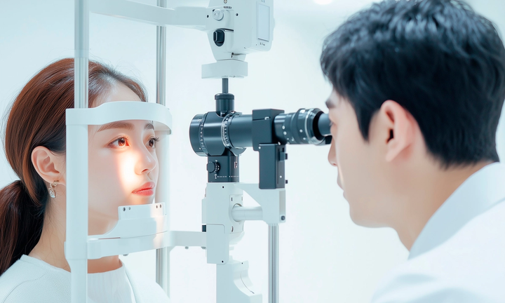
The examination before ICL measures the space the lens will occupy and the tissues next to it, in addition to the refraction and corneal measurements taken before any refractive procedure. The main items are listed here in brief. Each can be measured with more than one instrument, and clinics differ in which they use.
- Refraction, and whether it has been stable — the U.S. approval requires a change of no more than 0.5 dioptres over the preceding year.
- Anterior chamber depth — measured from the back of the cornea to the front of the natural lens. Where an instrument measures from the front surface of the cornea, corneal thickness is subtracted to arrive at that figure.
- The drainage angle — examined with a lens placed on the eye (gonioscopy). The approval requires an open angle, and the angle can become narrower after the lens is in place.
- Endothelial cell density — the cells lining the back of the cornea are counted (specular microscopy) and compared against a minimum that depends on age and anterior chamber depth.
- Measurements for choosing the lens size — including the horizontal width of the cornea and, where available, the dimensions of the space behind the iris. Lens size is chosen to help achieve an appropriate vault.
- Pupil size in dim light — a pupil that opens wider than the optical zone of the lens is associated with glare and halos at night.
- Eye pressure, and an examination of the retina — high short-sightedness carries its own retinal risk, which is assessed before any refractive surgery.
What each of these measurements means for an individual eye is a matter for the examining ophthalmologist.
Who is not a candidate for ICL?
Suitability rests on measurements of the front of the eye rather than on the cornea. In the United States the approval applies to adults aged 21 to 60 and specifies an anterior chamber depth of 3.00 mm or greater, measured from the back of the cornea to the front of the natural lens. The U.S. approval also covers defined ranges of short-sightedness, with correction from -3.0 to -15.0 dioptres and reduction from -15.0 to -20.0 dioptres. Approved ranges differ by regulator, so these figures describe the U.S. indication rather than a global rule. Published studies and selection criteria used outside the United States sometimes discuss lower anterior chamber depths, such as 2.80 mm, but the U.S. indication uses 3.00 mm. Endothelial cell density is assessed against a minimum appropriate to the person's age. Published studies of the procedure have commonly used 2,000 cells per square millimetre or above as an inclusion threshold, which indicates the range clinicians work with rather than a fixed clinical cut-off.
A prescription that is still changing rules the procedure out, as it does for laser surgery. In the United States the approval requires stability within 0.5 dioptres over the preceding year. Existing cataract, glaucoma, uveitis, and previous retinal detachment are assessed, as are autoimmune conditions and pregnancy.
Age matters in a way it does not for laser procedures. The natural lens is left in place, so an eye already developing cataract is generally directed elsewhere. How long LASIK results last covers the age-related changes that continue regardless of any refractive procedure.
Good to know
The measurements that decide ICL suitability are not the ones that decide it for laser surgery. An eye ruled out for one may still be assessed for the other, and the reverse is equally true. Neither result predicts the other, which is why the examination covers both sets of measurements.
Medical information
This article is general information about refractive surgery and is not medical advice. Individual results differ. Decisions about whether a procedure is appropriate, and about follow-up care, should be made with a qualified ophthalmologist after examination.